3D Corneal Mapping: Why Precision Matters (And Why We’re Obsessed With It)
Hey there, fellow vision enthusiasts! Ever wonder how your eye surgeon can transform a blurry world into crisp HD without even touching a scalpel? Spoiler: It starts with knowing your cornea like the back of their hand. And we’re not talking about a rough sketch here. At Liberty Laser Eye Center in Vienna, Virginia, 3D corneal mapping is our secret weapon—and today, we’re pulling back the curtain on why it’s a total game-changer.
Picture this: Your cornea isn’t just a smooth dome. It’s a complex, asymmetrical landscape with peaks, valleys, and subtle imperfections. Trying to correct nearsightedness, farsightedness, or astigmatism without mapping it first? That’s like navigating D.C. traffic blindfolded. Not ideal.
What Exactly Is 3D Corneal Mapping?
Think of it as GPS for your eyeballs. While old-school methods gave us a flat, 2D snapshot, modern mapping (using tech like Wavefront Analysis and Topography-Guided LASIK Surgery) creates a micron-level 3D model. We capture over 25,000 data points in seconds—no puff-of-air torture involved.
Why geek out over this? Because:
- Irregular astigmatism? Mapped.
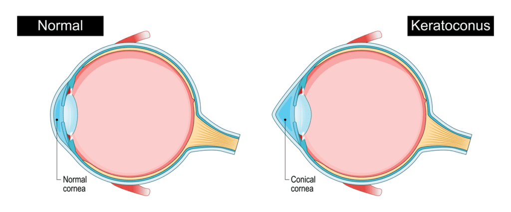
- Early keratoconus (that cone-shaped bulge)? Spotted before it’s a crisis.
- Dry eye hotspots? Flagged instantly.
Without this, laser eye surgery is like baking a cake without measuring cups. Sure, it might work… but do you feel lucky?
Precision Isn’t Just Fancy—It’s Non-Negotiable
Here’s the raw truth: Your cornea’s shape is as unique as your fingerprint. A “close enough” approach might leave you with glare, halos, or underwhelming vision. At Liberty Laser Eye Center, we refuse to roll those dice.
Consider Topography-Guided LASIK. Standard LASIK corrects basic glasses prescriptions. But if you’ve got quirky corneas? Topography-guided uses your 3D map to direct the laser exactly where it’s needed. Success rates soar, and night vision improves dramatically. IMO, it’s the difference between a flip phone and a smartphone.
Wavefront Analysis goes deeper, mapping how light scatters through your entire optical system. It’s why we catch sneaky issues even you didn’t notice. (FYI—this tech is standard in our D.C.-area clinic because “good enough” isn’t in our vocabulary.)
When Precision Saves Your Vision (Literally)
Keratoconus patients, this one’s for you. Early detection via 3D mapping is everything. Spot it soon enough, and Corneal Cross-Linking can stabilize your cornea. Miss it? You’re facing transplants or permanent haze.
Presbyopia warriors (yes, we see you squinting at menus), PresbyLASIK Surgery uses mapping to create a multifocal effect on your cornea. No more readers on your head!
And for those with thin corneas or dry eyes? Advanced PRK Surgery—guided by mapping—is often safer than LASIK. Recovery takes longer, but precision minimizes risks.
Here’s how mapping tackles common villains:
| Condition | How 3D Mapping Wins |
|---|---|
| Astigmatism | Finds hidden irregularities missed by standard tests |
| Keratoconus | Detects early thinning; guides Cross-Linking |
| Presbyopia | Designs custom “blended vision” for near AND distance |
| Dry Eye | Maps tear film breaks to avoid laser zones that could worsen symptoms |
Why Liberty Laser Eye Center? (Hint: We’re Laser-Focused)
Look, not all Lasik eye surgeons prioritize mapping. Some still use 2005 tech. But in our Vienna, VA clinic—just minutes from Washington DC—we treat your corneas like priceless art. Our best Lasik surgeons lean on mapping because:
- It cuts complication risks by up to 40% vs. non-mapped procedures.
- Results feel tailor-made. Goodbye, “20/20 with starbursts.”
- We screen out bad candidates. Not everyone qualifies for LASIK (shocking, right?), and mapping exposes deal-breakers fast.
Plus, our reviews don’t lie. Patients drive from Maryland and beyond because precision equals consistency.
“But What About Cost?” (Let’s Demystify)
We get it—laser eye surgery feels like a splurge. But here’s a not-so-secret fact: 3D mapping often makes surgery more affordable long-term. How? Fewer touch-ups. Zero “oops” moments. And hey, we offer financing because great vision shouldn’t require a trust fund.
Compare the “precision premium”:
- Standard LASIK: Corrects basic vision.
- Mapped LASIK/PRK: Corrects vision + hidden distortions + reduces side effects.
Which sounds like a smarter investment? 🙂
Your Game Plan: From Mapping to Marvelous Vision
Ready to explore eye surgery types? Here’s how we roll at Liberty:
- Comprehensive Mapping Session: 20 mins, zero discomfort. We’ll explain every hill and valley.
- Personalized Plan: LASIK? PRK? PresbyLASIK? We match you to the right surgery.
- Surgery Day: Most procedures take 10 mins per eye. Yes, really.
- Lasik Recovery: Most folks binge Netflix the next day. We’ll coach you through it.
Annual eye exams catch issues early. But if you’re Googling “Lasik doctors near me,” skip the guesswork. We’re the nearest precision-obsessed team in Northern Virginia.
Bottom Line: Don’t Trust Your Eyes to Amateurs
Sarcasm alert: If you want crooked vision correction, there’s a discount clinic next to a nail salon. But if crisp, clear sight matters? 3D mapping is non-negotiable. It’s why our success rate makes rivals sweat—and why D.C. professionals trust us with their most valuable asset.
So, nearsighted friends, astigmatism warriors, or presbyopia pioneers: Stop squinting at screens searching for “affordable laser eye surgery nearby.” Come see why precision isn’t just our policy—it’s our obsession.
Ready for eye maps that put Google Earth to shame? Contact Liberty Laser Eye Center in Vienna, VA today. Your corneas deserve it.
FAQs: Your Curiosities, Answered
Q: Does corneal mapping hurt?
A: Nope! It’s a quick, painless scan. You stare at a light, and we handle the rest.
Q: Can mapping help if I’ve been told I’m not a LASIK candidate?
A: Absolutely! We’ve cleared “non-candidates” using topography-guided PRK or Cross-Linking.
Q: How often should I get mapped?
A: Annually if you have keratoconus or dry eye. Otherwise, every 2 years unless symptoms change.
Q: Why choose Liberty over big-name chains?
A: Our Lasik eye surgeons in Vienna, VA aren’t factory workers. You get personalized care from start to finish—no cattle calls.
People Also Ask
Corneal topography is a highly accurate and non-invasive diagnostic tool used to map the surface curvature of the cornea. Its precision is essential for diagnosing conditions like keratoconus, planning refractive surgeries such as LASIK, and fitting contact lenses. Modern topographers, which use placido disc, slit-scanning, or Scheimpflug imaging technology, provide extremely detailed and quantitative maps of corneal shape, elevation, and power. Accuracy is typically within microns for elevation data. However, accuracy can be influenced by factors like patient cooperation, tear film quality, and operator skill. In a clinical setting, it is considered the gold standard for corneal shape analysis, providing reproducible and reliable data when performed correctly under standardized conditions.
Corneal topography is crucial because it provides a detailed, three-dimensional map of the cornea's surface curvature. This diagnostic tool is essential for detecting irregular astigmatism, keratoconus, and other corneal dystrophies that a standard eye exam might miss. It is a fundamental step in pre-operative planning for laser vision correction (like LASIK), as it ensures the cornea is healthy and suitable for surgery, thereby enhancing procedural safety and accuracy. Furthermore, it is vital for fitting specialized contact lenses and monitoring corneal changes over time. In essence, it offers an objective, non-invasive analysis that is critical for both diagnosis and the successful management of a wide range of ocular conditions.
Using a digital corneal topographer provides a significant advantage in precision and diagnostic capability. It creates a highly detailed, three-dimensional map of the cornea's surface, capturing thousands of data points to measure its shape, curvature, and elevation with micron-level accuracy. This is far superior to older manual methods. This detailed mapping is crucial for accurately diagnosing conditions like keratoconus, for planning refractive surgeries such as LASIK, and for achieving optimal fits for specialty contact lenses, including those for orthokeratology. The digital data allows for advanced analysis, trend tracking over time, and seamless integration with other diagnostic devices for a comprehensive view of ocular health.
The Pentacam, a rotating Scheimpflug camera system, offers significant advantages in comprehensive anterior segment analysis. Its primary benefit is providing a three-dimensional map of the cornea, delivering highly accurate measurements of corneal thickness, curvature, and elevation. This is crucial for diagnosing conditions like keratoconus and for planning refractive surgeries such as LASIK, where it helps screen for unsuitable candidates. The device also allows for detailed analysis of the anterior chamber, including depth and angle, which is vital for assessing glaucoma risk. Unlike older technologies, it directly measures the true corneal power, improving the accuracy of intraocular lens calculations for cataract surgery. This results in more predictable surgical outcomes and enhanced patient safety.
A corneal topography test is a non-invasive diagnostic procedure that creates a detailed three-dimensional map of the cornea's surface curvature. It is a standard tool in ophthalmology and optometry, primarily used for pre-operative screening for LASIK and other refractive surgeries to detect irregularities like keratoconus. The test also aids in fitting contact lenses, diagnosing corneal diseases, and evaluating the cornea post-surgery or post-injury. During the procedure, the patient focuses on a target while a computerized instrument projects a series of concentric rings onto the cornea, analyzing the reflected patterns to generate the topographic map. This provides critical data on corneal shape, power, and any astigmatism, which is essential for ensuring safe surgical outcomes and accurate vision correction.